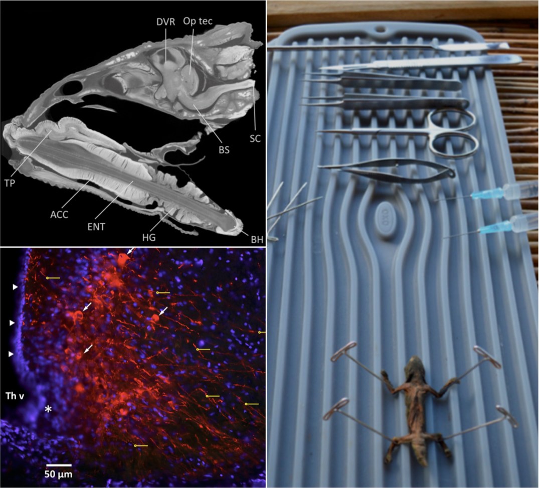
“Our understanding of the diversity and function in the vertebrate brain has been limited in part by the reliance upon laboratory facilities to preserve brain tissue under optimal conditions. In this study, we field-tested two standard laboratory-based techniques for brain preservation in an African biodiversity hotspot. We validated these protocols across multiple scales of analysis through cytoarchitectonic and immunohistochemical comparisons between field and laboratory-fixed tissue sets and diceCT imaging. In particular, diceCT images revealed excellent contrast of brain tissue structures, including myelinated and unmyelinated portions of the brain. Our protocol should serve as a malleable framework for researchers intending to explore the brains of poorly known and often inaccessible vertebrate species.”
– Project Leaders Daniel Hughes & Arshad Khan
