Featured Editorial Team: The Anatomical Record

unnamed document   unnamed document 1

“The ability to visualize structures in three-dimensional space will revolutionize how we can evaluate biomechanics in a wholly non-destructive approach. In addition, the possibility of algorithmic approaches (like those published recently by Dickinson and colleagues) will improve repeatability, and likely will help us save data collection time.”

– Adam Hartstone-Rose,

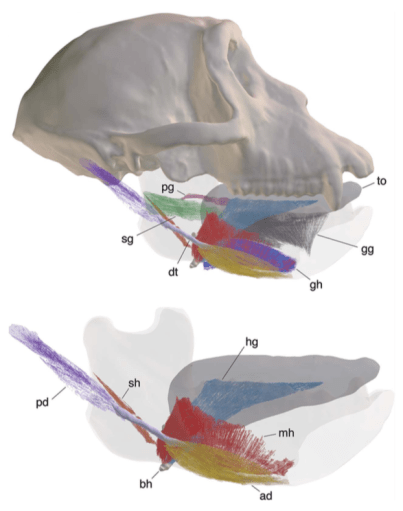
Traditional methods get paired with new imaging techniques to advance the study musculoskeletal biomechanics, featured in the February and March 2018 Special Issues of The Anatomical Record edited by Hartstone-Rose and his colleagues Sharlene Santana, Damiano Marchi, & Jeffrey T. Laitman.

New Publication: Assessment of the Hindlimb Membrane Musculature of Bats: Implications for Active Control of the Calcar

Screen Shot 2018-02-12 at 10.01.28 AM
3D diceCT models and histological sections through the calcar of (a) Myotis californicus, (b) Artibeus jamaicensis, (c,d) Molossus molasses. Specimens were stained with Lugol’s iodine for contrast-enhanced X-ray µCT imaging, subsequently destained by leaching in 70% EtOH, and re-stained for histological sectioning using Modified Mayer’s Hematoxylin and Mallory triple connective tissue stains. Abbreviations: Ca, calcar; m.A, additional muscle in M. molossus; m.CC, m. calcaneocutaneous; m.D, m. depressor ossis styliformis; m. DP, m. depressor ossis styliformis profundus; m.DS, m. depressor ossis styliformis superficialis.

“Exploring the detailed muscular anatomy of very small mammals is difficult to do using dissection alone since often the details are damaged before they can be observed properly. Here, we used diceCT to learn more about the musculature associated with the bat calcar — a skeletal novelty found in bat feet. DiceCT combined with standard histology revealed anatomical variation among the calcar musculature of different bat species that quite possibly has functional implications. This could mean that the calcar has functionally diversified among bats. DiceCT is an extremely useful tool for revealing previously-unknown anatomical diversity, especially in small animals.”


Lead author, Kathryn Stanchak (@)

See more of this research at the Bat Cave on Twitter, the Santana Lab website, and read the the pub at The Anatomical Record!

New Publication: Specialized specialists and the narrow niche fallacy: a tale of scale-feeding fishes


“Lepidophagous fishes, which subsist by picking scales off other fishes, have evolved independently over 30 times. Given the seemingly specialized nature of this dietary niche, we asked the question: are all scale-feeding fishes built in a similar fashion? We used microCT to measure the feeding anatomy in a host of museum specimens, coupled with diceCT to visualize jaw musculature without marring priceless specimens with dissections. Despite living in similar habitats and feeding on presumably similar prey, lepidophagous taxa do not converge on a singular morphotype; rather, there are many ways to be a scale-feeding fish.”

– Lead author, Matt Kolmann (@)

Follow Dr. Kolmann on Twitter, and head over to Royal Society Open Science to read the open access pub!

New Publication: Dynamic Musculoskeletal Functional Morphology: Integrating diceCT and XROMM

Screen Shot 2018-02-05 at 11.46.56 AM
Select macaque hyolingual muscles. (Top) Lateral view of cranium, mandible (transparent), basihyoid, and select hyolingual muscles; (Bottom) Same view as top, showing only the muscles that are hypothesized to produce hyoid elevation. Abbreviations (color): ad, anterior digastric (yellow); bh, basihyoid (tan); gg, genioglossus (dark gray); gh, geniohyoid (blue); hg, hyoglossus (light blue); mh, mylohyoid (red); pd, posterior digastric (purple); pg, palatoglossus (pink); sg, styloglossus (green); sh, stylohyoid (orange); to, tongue (light gray). (The kinematics of these muscles were reconstructed using a combination of XROMM and FMM.)

“Studying muscle functional morphology is often easier said than done because definitively determining muscle function requires measuring many morphological and physiological parameters simultaneously. To advance our studies of the primate hyolingual apparatus, which is composed of dozens of muscles, we developed a pipeline to integrate diceCT and EMG with X-ray Reconstruction of Moving Morphology (XROMM, Brainerd et al. 2010). XROMM obtains the high spatiotemporal resolution kinematics necessary to analyze the three dimensional complexity of hyolingual movement, and incorporating diceCT provides a new method for confirming EMG electrode location and improves the accuracy of muscle and fiber length and orientation measurements. Together, these methods will help scientists to determine how organisms navigate the many ways of tuning organismal performance through musculoskeletal design.”


-Lead Author, Courtney Orsbon (@)

Find more methods integration at the Ross Lab website, and read the pub at The Anatomical Record!