Welcome!

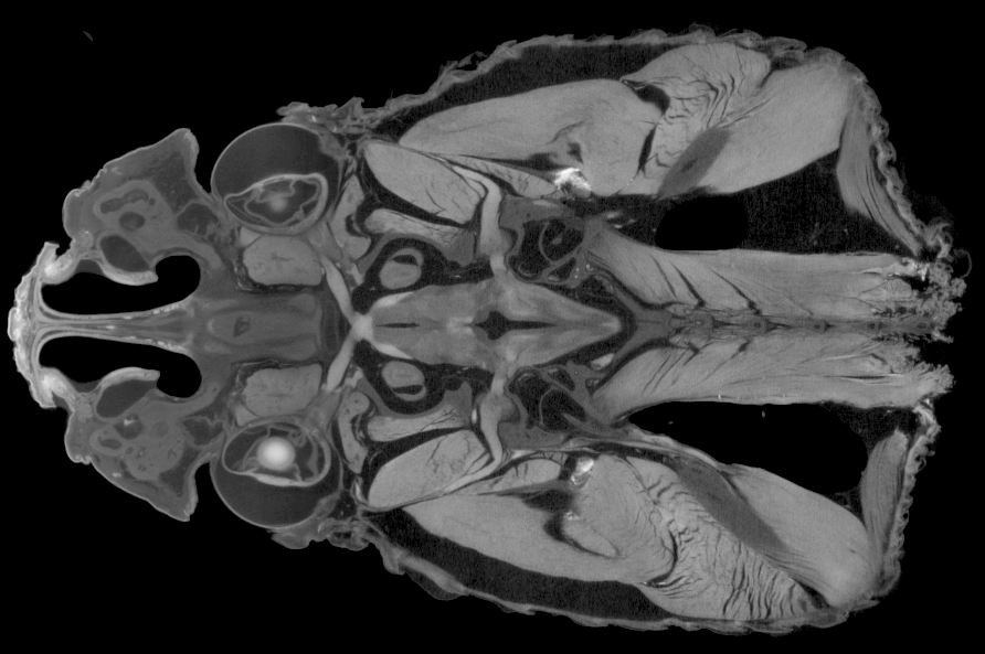
Welcome to the online home of Diffusible Iodine-based Contrast-enhanced Computed Tomography.

Our mission is to provide digital resources for the diceCT community and to connect interested researchers with contrast-enhanced imaging veterans. Watch this space and @diceCT for updates on new publications, tips & tricks, and diceCT-related events.

New Content

The Austin Working Group | 2015 SVP Workshop | 2016 ICVM Symposium | Pubs

Crotalus_atrox_frontal
Computed tomography section through the head of a western diamondback rattlesnake in frontal view, prepared using contrast enhancement. Soft tissues including epithelia, glands, cranial muscles, and myelinated as well as non-myelinated nervous tissues can be readily visualized alongside bony structures using diceCT techniques. (Thanks to Dr. Arshad Khan & The University of Texas, El Paso for securing this specimen.)
supported

Imaging Technology Symposium—Summer 2019!

image001

Advances in CT Technologies for LifeScience and Natural History Museum Collections

The Museum für Naturkunde and YXLON are partnering to deliver an Imaging Technology Symposium, June 12–13th 2019 in Berlin!

The symposium will bring together users of CT technology from biology, morphology, paleontology, zoology, and related areas to share their expertise and knowledge about exciting developments in biological imaging. A hands-on session by YXLON with their FF35 CT scanner (on June 14th) will follow the 2-day symposium.

The scientific program will consist of lectures from international scientists in the field as well as practically focused, hands-on and workshop sessions.

Interested? See more and register at: https://www.yxlon.com/news-events/events/events/2019-imaging-technology-symposium

DiceCT Centennial!

dicect_centennial

30 months after the Journal of Anatomy published the eponymous and open access diceCT review, we have crossed the 100 citations threshold!

The Austin Working Group is thrilled to see how a community of passionate, creative, and clever researchers—spanning the globe—have come together around contrast-enhanced imaging tools and diceCT in such a short time!

We’re particularly proud of the open nature of the community, sharing tips and tricks, successes and failures with each other and with newcomers to the field. You have helped to catapult the success of 3D soft-tissue imaging from a comparatively low-resolution, expensive diagnostic tool to an accessible, next-generation research visualization platform.

Here’s to another excellent 30 months of stellar research and remarkable visuals! 🍾🎉

New Publication: The Utility of DiceCT Imaging for High-Throughput Comparative Neuroanatomical Studies

KargerBBE_GignacKley
DiceCT facilitates the rapid visualization of both external and internal brain anatomy in vertebrates – alongside the intact bones of the skull and the complete, undisturbed pathways of peripheral nerves, up to and including the target organs that they innervate. This approach allows for the digital extraction of vertebrate brains across 10,000-fold ranges in specimen size and at micron-scale resolutions.

” Like the current ‘gold standard’ of magnetic resonance imaging, diceCT does not require physical dissection and can differentiate between the lipid content of myelinated versus nonmyelinated tissues, thereby offering great potential for neuroanatomical studies. Within the brain diceCT distinguishes myelinated fiber tracts from unmyelinated cortices, nuclei, and ganglia and allows 3D visualization of their anatomical interrelationships at previously unrealized spatial scales. In our open access study,  we demonstrate the transformative potential of diceCT for developing high-resolution neuroanatomical datasets and describe best practices for imaging large numbers of specimens for broad evolutionary studies across vertebrates.”

– lead author, Paul Gignac (@)

Head over to Brain, Behavior and Evolution to read this open access pub and find more excellent neuroscience from the Karger workshop special edition, From Fossils to Function: Integrative and Taxonomically Inclusive Approaches to Vertebrate Evolutionary Neuroscience

New Publication: Assessing Soft-Tissue Shrinkage Estimates in Museum Specimens Imaged With Diffusible Iodine-based Contrast-Enhanced Computed Tomography

Pallas’s long-tongued bat
Mid-sagittal section of museum specimen of Glossophaga soricina (Pallas’s long-tongued bat) head after three weeks in I2KI stain demonstrating complete penetration after only several weeks in stain. The black space surrounding the brain shows the degree of shrinkage. Anatomical structures are outlined for orientation. Scale bar = 4 mm.

DiceCT allows visualization of organismal soft-tissue cheaply and non-destructively, thus giving comparative biologists a new toolkit for assessing morphological variation. Comparative morphologists primarily use museum collections to visualize features across a wide range of species, but the consequences of preparation and storage are not well understood. We report soft-tissue shrinkage in the brains and eyes of five bat species from museum collections and compare this to shrinkage found in specimens of six freshly-collected species. Although the magnitude of shrinkage in the museum specimens did not increase over four weeks of stain time in iodine, the brains and eyes of museum specimens shrank considerably prior to placement in iodine in comparison with field-collected specimens. While the cause of shrinkage in these specimens remains unknown, we caution against study designs that combine fresh and museum specimens.

Lead author, Brandon Hedrick (@)

Read more about using diceCT with museum specimens at Microscopy & Microanalysis!

New Publication: Cephalic muscle development in the Australian lungfish, Neoceratodus forsteri

Fig. 4 Levatores arcuum branchialium LZW 29Oct17 copy
Neoceratodus forsteri, develoment of branchial arch muscles. Top row: Sagittal sections; Levatores arcuum branchialium I-V (LAB I-V) attaching to ceratobranchiales (CB) I-V (anterior is left; stage 52/53) . Bottom row: Branchial arch muscles in in a juvenile N. forsteri, diceCT images, sagittal images; LAB (I-V) (anterior is left). See publication for details of histological staining; scale bars are 1 mm.

“Our current understanding of development and evolution of head and neck musculature in vertebrates is often based on studies in a few model organisms that might or might not be at relevant positions on a phylogenetic tree to highlight key changes from a primitive to a derived character. Lungfishes, like the Australian Lungfish, are at one such relevant position as their anatomy and ontogeny can help us to understand the changes that occurred during thewater to land transition. Methods, like diceCT, that allow us to analyze in detail the anatomy of species without destructive dissections are of increasing value as they not only to reduce the amount of specimens needed to investigate but also allow the 3D visualization of complex structures, which in turn enables us to make more precise predictions about functional changes due to differences of muscles attachments at different stages of development. Comparisons of developmental changes with differences observed during the evolution of vertebrate species will then allow us to identify highly conserved or less restricted mechanisms that play a role during the evolution of diverse species from fish to humans.”

–authors, Alice ClementJanine Ziermann (@)

Read more about Australian lungfish at the Journal of Morphology and see more work like this on ResearchGate and at PLOS ONE!


New Publication: Vomeronasal and Olfactory Structures in Bats Revealed by DiceCT Clarify Genetic Evidence of Function

3D Bat Vomeronasal Organs
Coronal sections of the posterior region of the nasal cavity comparing diceCT scans and 3D reconstructions in bats (frontoturbinal is light green; interturbinals are dark green, ethmoturbinal I is yellow; ethmoturbinal II is light blue; and ethmoturbinal III is teal; see publication for abbreviations.)

“Bats demonstrate one of the most impressive stories of independent loss of the vomeronasal organ, a specialized nasal pheromone-sensing system in mammals. We were surprised to learn of a loss-of-function mutation in a vomeronasal-specific gene within a clade of Caribbean nectar-feeding bats, as many of their mainland relatives maintain function of the gene and organ—but the morphology was not known for this clade. DiceCT permitted us to peek inside the heads of these bats and characterize the nasal soft tissues, including the first 3D reconstruction of a vomeronasal organ, a structure only thought visible through histology. We discovered that Caribbean nectarivorous bats indeed have lost or reduced the vomeronasal organ and possess more elaborate olfactory turbinals. Complete loss of morphology likely occurred prior to complete genetic loss of function revealing a deeper understanding of the process of vestigalization.”

– co-lead authors, Laurel Yohe (@) & Simone Hoffmann (@)

Head over to Frontiers in Neuroanatomy to read the pub and see more research on Twitter!

Featured Editorial Team: The Anatomical Record

unnamed document   unnamed document 1

“The ability to visualize structures in three-dimensional space will revolutionize how we can evaluate biomechanics in a wholly non-destructive approach. In addition, the possibility of algorithmic approaches (like those published recently by Dickinson and colleagues) will improve repeatability, and likely will help us save data collection time.”

– Adam Hartstone-Rose,

Traditional methods get paired with new imaging techniques to advance the study musculoskeletal biomechanics, featured in the February and March 2018 Special Issues of The Anatomical Record edited by Hartstone-Rose and his colleagues Sharlene Santana, Damiano Marchi, & Jeffrey T. Laitman.

New Publication: Assessment of the Hindlimb Membrane Musculature of Bats: Implications for Active Control of the Calcar

Screen Shot 2018-02-12 at 10.01.28 AM
3D diceCT models and histological sections through the calcar of (a) Myotis californicus, (b) Artibeus jamaicensis, (c,d) Molossus molasses. Specimens were stained with Lugol’s iodine for contrast-enhanced X-ray µCT imaging, subsequently destained by leaching in 70% EtOH, and re-stained for histological sectioning using Modified Mayer’s Hematoxylin and Mallory triple connective tissue stains. Abbreviations: Ca, calcar; m.A, additional muscle in M. molossus; m.CC, m. calcaneocutaneous; m.D, m. depressor ossis styliformis; m. DP, m. depressor ossis styliformis profundus; m.DS, m. depressor ossis styliformis superficialis.

“Exploring the detailed muscular anatomy of very small mammals is difficult to do using dissection alone since often the details are damaged before they can be observed properly. Here, we used diceCT to learn more about the musculature associated with the bat calcar — a skeletal novelty found in bat feet. DiceCT combined with standard histology revealed anatomical variation among the calcar musculature of different bat species that quite possibly has functional implications. This could mean that the calcar has functionally diversified among bats. DiceCT is an extremely useful tool for revealing previously-unknown anatomical diversity, especially in small animals.”


Lead author, Kathryn Stanchak (@)

See more of this research at the Bat Cave on Twitter, the Santana Lab website, and read the the pub at The Anatomical Record!

New Publication: Specialized specialists and the narrow niche fallacy: a tale of scale-feeding fishes


“Lepidophagous fishes, which subsist by picking scales off other fishes, have evolved independently over 30 times. Given the seemingly specialized nature of this dietary niche, we asked the question: are all scale-feeding fishes built in a similar fashion? We used microCT to measure the feeding anatomy in a host of museum specimens, coupled with diceCT to visualize jaw musculature without marring priceless specimens with dissections. Despite living in similar habitats and feeding on presumably similar prey, lepidophagous taxa do not converge on a singular morphotype; rather, there are many ways to be a scale-feeding fish.”

– Lead author, Matt Kolmann (@)

Follow Dr. Kolmann on Twitter, and head over to Royal Society Open Science to read the open access pub!

New Publication: Dynamic Musculoskeletal Functional Morphology: Integrating diceCT and XROMM

Screen Shot 2018-02-05 at 11.46.56 AM
Select macaque hyolingual muscles. (Top) Lateral view of cranium, mandible (transparent), basihyoid, and select hyolingual muscles; (Bottom) Same view as top, showing only the muscles that are hypothesized to produce hyoid elevation. Abbreviations (color): ad, anterior digastric (yellow); bh, basihyoid (tan); gg, genioglossus (dark gray); gh, geniohyoid (blue); hg, hyoglossus (light blue); mh, mylohyoid (red); pd, posterior digastric (purple); pg, palatoglossus (pink); sg, styloglossus (green); sh, stylohyoid (orange); to, tongue (light gray). (The kinematics of these muscles were reconstructed using a combination of XROMM and FMM.)

“Studying muscle functional morphology is often easier said than done because definitively determining muscle function requires measuring many morphological and physiological parameters simultaneously. To advance our studies of the primate hyolingual apparatus, which is composed of dozens of muscles, we developed a pipeline to integrate diceCT and EMG with X-ray Reconstruction of Moving Morphology (XROMM, Brainerd et al. 2010). XROMM obtains the high spatiotemporal resolution kinematics necessary to analyze the three dimensional complexity of hyolingual movement, and incorporating diceCT provides a new method for confirming EMG electrode location and improves the accuracy of muscle and fiber length and orientation measurements. Together, these methods will help scientists to determine how organisms navigate the many ways of tuning organismal performance through musculoskeletal design.”


-Lead Author, Courtney Orsbon (@)

Find more methods integration at the Ross Lab website, and read the pub at The Anatomical Record!