Thanks to the Society of Integrative and Comparative Biology‘s Division of Vertebrate Morphology for a shoutout to the Austin Working Group in the Fall 2015 Society Newsletter!

Thanks to the Society of Integrative and Comparative Biology‘s Division of Vertebrate Morphology for a shoutout to the Austin Working Group in the Fall 2015 Society Newsletter!
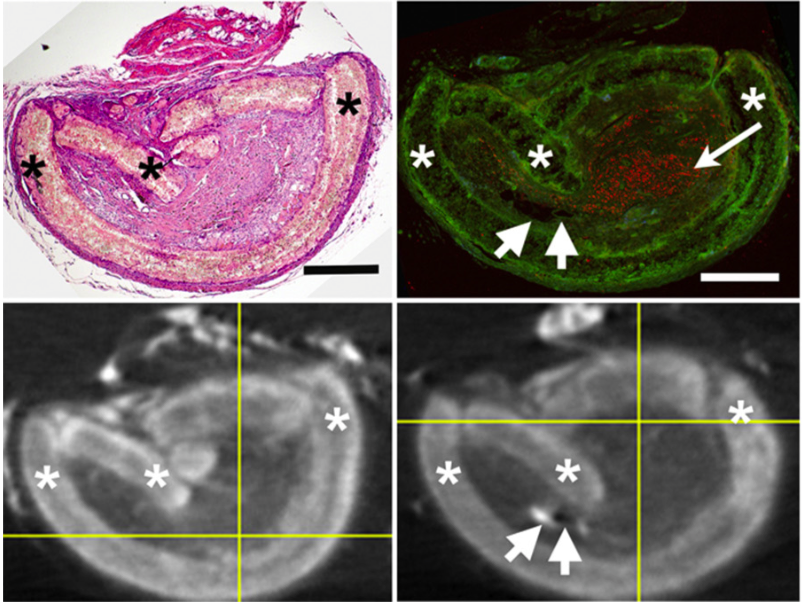
“In pre-clinical studies, biomaterial implants with densities similar to tissues cannot be imaged via standard μCT after removal from the animal. These ex vivo implants then present investigative challenges with histological analyses because of implant size, and critical information can be lost. We used iodine-based contrast-enhanced μCT imaging to visualize polymeric biomaterial nerve guides used to repair injury gaps in rodent peripheral nerves, followed by rapid removal of the iodine, and showed that these treatments did not interfere with histological or immunohistological analyses. This increases the information that can be obtained from one set of animals, allows novel comparisons and thus could help researchers studying any soft tissue or tissue-density biomaterial implant.”
–Author Sarah Pixley @
Head over to the Journal of Neuroscience Methods to read the pub!

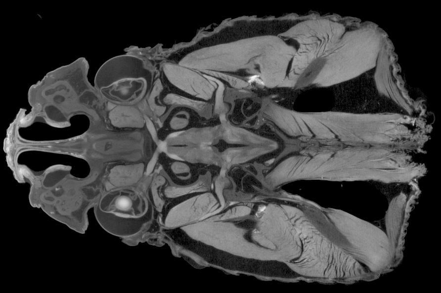
“Traditionally, taxonomic descriptions have relied on drawings and photographs to record morphological information that distinguishes different species. Modern 3-D imaging technology and particularly X-ray microtomography provides not only a third dimension to the pictures, but also the means for sharing the information widely. We have thought of creating a cybertype-enhanced description of a new millipede from Spain, dubbed Ommatoiulus avatar in recognition of its digital alter ego. This is the first new species to be described with the aid of detailed 3-D images of the actual type specimens and to be published with its cybertypes as an open-access resource.“
–Lead author Nesrine Akkari
Welcome to the online home of Diffusible Iodine-based Contrast-enhanced Computed Tomography.
Our mission is to provide digital resources for the diceCT community and to connect interested researchers with contrast-enhanced imaging veterans. Watch this space and @diceCT for updates on new publications, tips & tricks, and diceCT-related events.
The Austin Working Group | 2015 SVP Workshop | 2016 ICVM Symposium | Pubs