A diceCT visualized ratsnake head and brain is a finalist in the 2016 Royal Society of London Scientific Photography Competition! This piece and many other contributions of remarkable scientific artwork are now part of a display at the Royal Society of London Online.
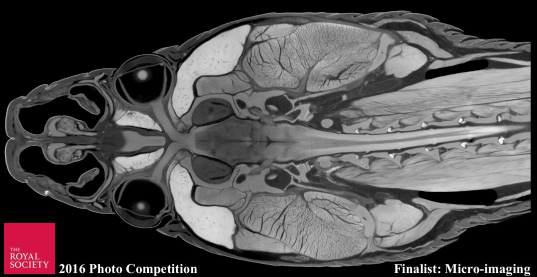
“The cranial anatomy of a diceCT imaged ratsnake in frontal view” by Nathan Kley and Paul Gignac. Ratsnake (genus Pantherophis) head in frontal view, prepared using diffusible iodine-based contrast enhanced computed tomography (diceCT) imaging, in which an iodine staining solution acts as a contrast agent that non-destructively renders soft tissues visible in X-ray micro-CT scans. The specimen was imaged using a GE Phoenix v | tome | x micro-CT scanner at 29.5 microns. Soft tissues including the brain and its internal structures, spinal cord, optic chiasm, roots and branches of cranial nerves, jaw and neck muscles, glands, nasal epithelia, cranial bones, and integument can all be clearly visualized simultaneously. (Image contrast and brightness were modified to render the background fully black and the neck was cropped to fit the image frame.)
The competition celebrates the power of photography to communicate science and the role images play in making science more accessible to a wide audience. To view featured photos at the Open House exhibition (9/17–9/18/2016), visit The Royal Society of London.