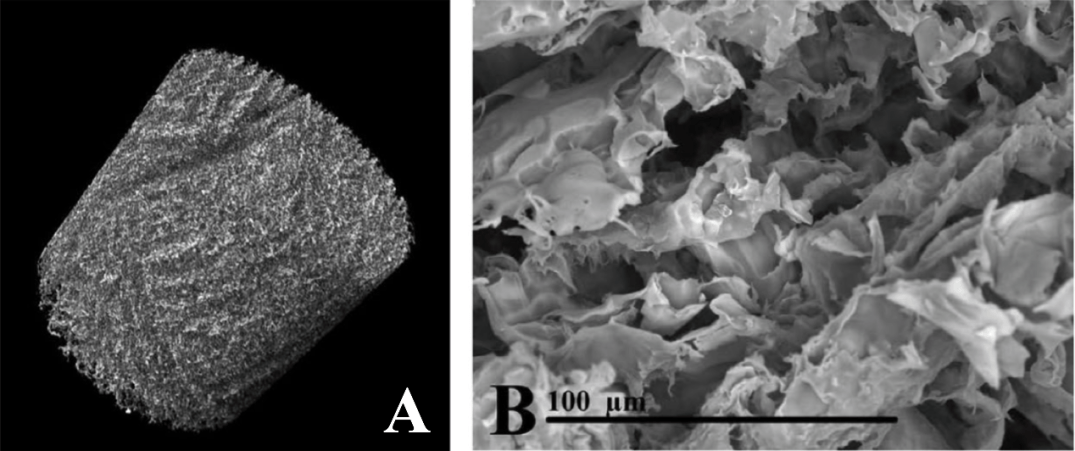
3-D rendering based on a μCT scan (A) alongside an SEM micrograph (B; 1000x magnification) of a biopolymer composite stained with 1.5 wt.% iodine and fixed with HMDS.
“μCT scanning of low density porous polymer scaffolds leads to insufficient imaging ability due to poor contrast. In order to enhance their X-ray absorption, one might trial several approaches using contrast agents. This methods paper is aimed at guiding scientists in applying the most appropriate contrast enhancement method for CT imaging of such materials, by reporting both the positive and the negative results. Iodine staining coupled with chemical drying was the most efficient approach, allowing for contrast enhancement, morphology preservation, and high resolution all at once.”
Two dozen speakers are scheduled to present in our diceCT symposium at ICVM in 2016.
Diffusible iodine-based contrast-enhanced computed tomography (diceCT) and related imaging techniques for research in evolutionary morphology
Organizers: P. M. Gignac1*, A. N. Herdina2, N. J. Kley3, A. Morhardt4, J. A. Clarke5, and M. Colbert5
The ability to visualize hard tissues (e.g., bone, dentine, enamel) rapidly in three dimensions using X-ray computed tomography (CT) has been one of the most important advancements in the field of vertebrate morphology in the last half-century. Until recently, however, comparably valuable advances in soft-tissue imaging have been difficult to realize fully due to the inherently low X-ray absorption of non-mineralized structures. Pioneering work in this area has demonstrated that Lugol’s iodine (I2KI) is a highly effective contrast agent for rapidly differentiating many types of soft tissues (e.g., epithelial, muscular, and neural structures) in micro computed tomography (μCT) images. Vertebrate morphologists have become a driving force in advancing this technique and utilizing the remarkable data generated from it to reconstruct phenotypes and functional anatomy in three and four dimensions.
Given the broad potential for iodine-enhanced CT imaging to become a major tool for soft-tissue reconstruction in vertebrate morphology, we will hold a symposium at the 2016 ICVM meeting to exhibit the wide range of taxa and questions that can be examined using these approaches. Through our symposium, Diffusible iodine-based contrast-enhanced computed tomography (diceCT) and related imaging techniques for research in evolutionary morphology, we will feature the newest and ongoing applications of contrast-enhanced three-dimensional (3-D) imaging already being undertaken by researchers within the International Society of Vertebrate Morphology. We further propose to hold a student-focused, combined poster session with the Hartstone-Rose and Marchi symposium on muscle functional morphology, to bridge the related techniques of our respective presenters. Our goal is to spur the further adoption of these methods by vertebrate morphologists. We will achieve this by (1) highlighting recent methodological advances in contrast-enhanced CT and μCT imaging, (2) demonstrating active research that integrates diceCT and related imaging techniques into toolkits addressing macroevolutionary questions, and (3) generating discussions of future directions and the long-term place for contrast-enhanced imaging in the study of extant vertebrates. We have assembled a diverse group of speakers who have enthusiastically agreed to participate in this symposium. They include well-established researchers, emerging early-career scientists, and graduate students in the fields of functional morphology, biomechanics, phenotypic integration, and vertebrate paleontology, whose academic contributions have already brought important insights to evolutionary biology. Demonstration of the high-level inferences that can be garnered with diceCT will spur collaborations among labs already exploring this powerful new tool with those who are considering how to apply it to their own research questions.
Poster Symposium
Green — Physiological examination of ratite orthopedic disorders and soft- tissue visualization via micro-CT
Herrel — Contrast-enhanced versus phase-contrast imaging: costs and benefits of different methods
Holliday — DiceCT and its applications for understanding the reptile musculoskeletal system
Introduction & Neurological Visualization
Gignac — DiceCTing the future: new horizons for 3-D visualization of vertebrate morphology (30 min)
Weisbecker — Using the STABILITY protocol prior to IKI-staining to provide the first accurate, in situ quantification of mammalian brain proportion scaling using marsupials (15 min)
Watanabe — Mind the Gap: ontogenetic shape differences between brains and endocasts in archosaurs (15 min)
Hughes — Incorporating diceCT into multi-scale structural studies of the brain for highly divergent lineages of acrodont Lizards: validation of preservation methods conducted in the field (15 min)
Functional Morphology
Gold — Applying diceCT to PET: new tools for correlating morphology to function in living animals (15 min)
Charles — Musculoskeletal modelling and simulations of the mouse hind limb during locomotion: the role of high-resolution scanning and contrast Imaging (15 min)
Lautenschlager — The evolution of the mammalian jaw adductor musculature – inferences from soft-tissue imaging of extant taxa (15 min)
Cox — Masticatory muscle anatomy of African mole-rats revealed by diceCT (15 min)
Orsbon— Integration of diceCT with XROMM and fluoromicrometry enhances functional morphology and biomechanics research: a case study of the macaque (Mammalia: Primates) feeding apparatus (15 min)
Descriptive Anatomy
Stanley —Contrast-enhanced CT provides insight into amphibian lingual morphology (15 min)
Pardo —Studying metamorphosis of the cranial musculoskeletal system in the axolotl using contrast-mCT (15 min)
Porro —Contrast-enhanced micro-CT imaging of fish and frogs: digital dissections and biomechanical applications (15 min)
Yohe —The curious case of the vomeronasal organ in bats: genetics asks questions only anatomy can answer (15 min)
Vander Linden —Comparative morphology of bat cranial muscles using contrast enhanced micro-CT imaging (15 min)
Advancements and Infrastructure for Contrast-enhanced Imaging Techniques
Herdina — Advantages and difficulties of alcoholic iodine staining for correlative 2-D and 3-D micro-CT imaging and histomorphology in bat developmental studies (15 min)
Mahlow — DiceCT and the staining of old museum specimens, exemplified by the analysis of venom glands in viperid snakes (15 min)
Morhardt — Diffusible iodine-based contrast-enhancement of large, post-embryonic, intact vertebrates for CT scanning: staining, destaining, and long-term storage (15 min)
Li — An evaluation of the efficacy and mechanism of contrast-enhanced X-ray computed tomography for avian cranial material utilizing iodine through experimental and simulation approaches (15 min)
Starck —The publishing and archiving of microscopic anatomy (15 min)
1*Department of Anatomy and Cell Biology, Oklahoma State University Center for Health Sciences, Tulsa, OK, USA (corresponding organizer: paul.gignac@okstate.edu)
2Department of Theoretical Biology, University of Vienna, Vienna, Austria
3Department of Anatomical Sciences, Stony Brook University, Stony Brook, NY, USA
4Department of Biomedical Sciences, Ohio University, Athens, OH, USA
5Jackson School of Geosciences, The University of Texas at Austin, Austin, TX, USA
Cover: An eclectus parrot (Eclectus roratus) stained by C. M. Holliday (University of Missouri) and micro-CT scanned by L. M. Witmer (Ohio University) according to the diceCT protocol presented by Gignac et al. (pp. 889–909) that reveals unprecedented soft-tissue detail in CT scanned specimens.
“Use of iodine as a contrast agent for computed tomography visualizations has been a growing area of research for the last several years. In this open access study we provide a summary of the diceCT literature and synthesize the elements of successful approaches: we analyze factors that govern success at each step of the diceCT imaging pipeline; we recommend a standard for reporting details of specimen preparation, storage, and imaging that will be pivotal to the repeatability and broader adoption of diceCT going forward; and we discuss cutting-edge applications and future directions for diceCT techniques. We anticipate robust growth within the diceCT community and the realization of new horizons in contrast-enhanced imaging—and it is our explicit purpose to spur the adoption of this relatively nascent approach by the morphology community as a whole.”
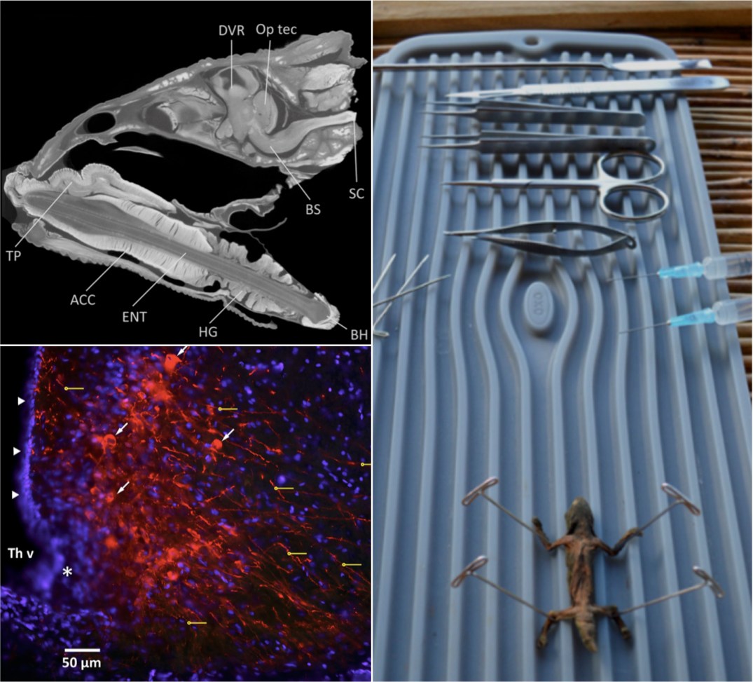
Multi-modal brain imaging from reptile specimens capture and fixed under remote field conditions (right) without cold-storage capabilities: cranial gross anatomy of male Trioceros johnstoni imaged using diceCT imaging (top left), and cellular-level, neural networks imaged from the same taxon (adjacent to the third ventricle), using a tyrosine hydroxylase immunoreactivity (-ir) stain (TH; red) with DAPI fluorescent counterstain (blue) (bottom left). (See manuscript for abbreviations.)
“Our understanding of the diversity and function in the vertebrate brain has been limited in part by the reliance upon laboratory facilities to preserve brain tissue under optimal conditions. In this study, we field-tested two standard laboratory-based techniques for brain preservation in an African biodiversity hotspot. We validated these protocols across multiple scales of analysis through cytoarchitectonic and immunohistochemical comparisons between field and laboratory-fixed tissue sets and diceCT imaging. In particular, diceCT images revealed excellent contrast of brain tissue structures, including myelinated and unmyelinated portions of the brain. Our protocol should serve as a malleable framework for researchers intending to explore the brains of poorly known and often inaccessible vertebrate species.”
Grayscale diceCT image of a European starling head in parasagittal view with a Positron Emission Tomography heat map superimposed. Warmer colors indicate metabolically active regions within the eye (left-most) and brain (right- and top-most). The anterior Wulst is encircled in the forebrain, indicating the highest levels of metabolic activity during flight are in this region.
“The morphological adaptations that led to flight in dinosaurs are understood, but the accompanying neuroanatomical changes are still elusive. We used Positron Emission Tomography scanning and diceCT to identify the parts of the brain used during flight in starlings. We found that the anterior Wulst and entopallium are the most used, probably creating a short-term conflict alert system that connects visual and somatosensory input to adjust flight paths. This gives us our first insight into how birds use their brains to fly and what that could mean for the evolution of volancy in theropod dinosaurs.”
Model and experimental data show the temporal and spatial profile (zones 1, 2, and 3) of iodine concentration. By recalibration of staining duration, a constant flux at the boundary condition in the model is generally expected to be met by maintaining the solution concentration in a certain level.
“There has been very limited data available to inform detailed protocols for staining large specimens using diceCT. In our study, we systematically assessed the efficacy and mechanism of diffusion-based contrast-enhanced X-ray Computed Tomography (CT) with serial experiments and the validation of numerical modeling. We have applied a Diffusion-Sorption model to explain the CT contrast increasing pattern within the cranial tissues of a goose specimen over a long staining duration. We identified attributes of different tissues that affect the effective diffusion rate and staining efficacy, including partition coefficient, bulk density and tissue porosity. Based on our results, specific protocols—customized by tissue size and type—can be designed for diceCT to maximize visualization of these soft tissues contrasts.”
Comparisons of MRI to diceCT mouse brains (left), showing displacement heat maps (right). Gray areas in the heat map indicate regions of large differences (>0.5 mm) either due to extreme shrinkage or difference in segmentation.
“Phenotypic screens for brain defects traditionally used either histology or high-resolution magnetic resonance imaging (MRI) to look for structural abnormalities. The former is tedious and slow, while the latter is expensive. By coupling the excellent contrast offered by the iodine with the tissue preserving properties of hydrogel, we demonstrate that whole mouse brains can be effectively and rapidly imaged by μCT at a fraction of a cost of high-resolution MRI. Our methodology is best suited for rapid phenotyping applications where large numbers of samples need to be screened for gross differences in the overall brain shape using computational morphometric approaches.”
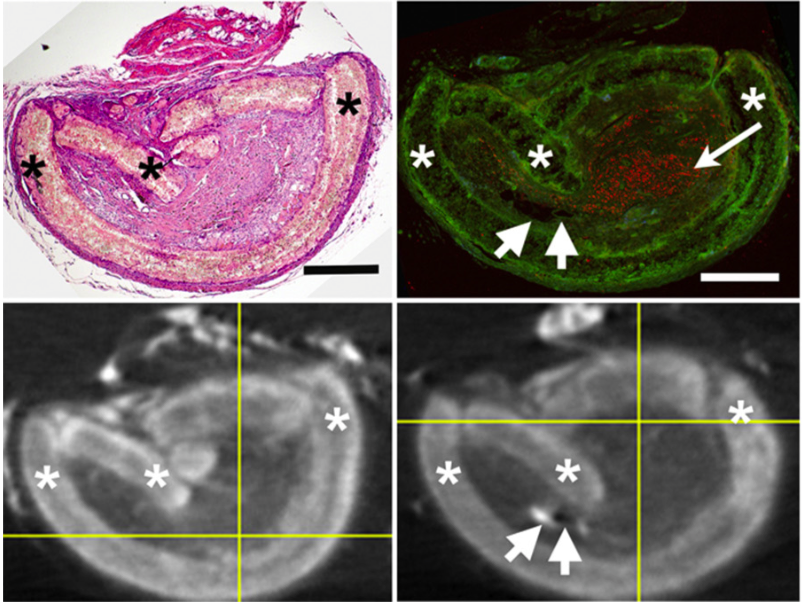
H&E stained section (top left) matched to the corresponding axial μCT image (bottom left) and a section stained with anti-NF (top right) matched to the axial μCT image (bottom right). Asterisks label nerve conduits. The long arrow points to a group of anti-NF stained axons, and short arrows point to a small pocket of fat (left) and a large blood vessel (right). Scale bars = 0.5 mm.
“In pre-clinical studies, biomaterial implants with densities similar to tissues cannot be imaged via standard μCT after removal from the animal. These ex vivo implants then present investigative challenges with histological analyses because of implant size, and critical information can be lost. We used iodine-based contrast-enhanced μCT imaging to visualize polymeric biomaterial nerve guides used to repair injury gaps in rodent peripheral nerves, followed by rapid removal of the iodine, and showed that these treatments did not interfere with histological or immunohistological analyses. This increases the information that can be obtained from one set of animals, allows novel comparisons and thus could help researchers studying any soft tissue or tissue-density biomaterial implant.”
3-D virtual dissection of the holotype depicting the gonopods in situ. (A) Oblique transverse cut showing attached musculature; (B) Parasagittal cut showing musculature and other anatomical structures
“Traditionally, taxonomic descriptions have relied on drawings and photographs to record morphological information that distinguishes different species. Modern 3-D imaging technology and particularly X-ray microtomography provides not only a third dimension to the pictures, but also the means for sharing the information widely. We have thought of creating a cybertype-enhanced description of a new millipede from Spain, dubbed Ommatoiulus avatar in recognition of its digital alter ego. This is the first new species to be described with the aid of detailed 3-D images of the actual type specimens and to be published with its cybertypes as an open-access resource.“